|
|
Histo-Pathological Image Analysis | Automated analysis of histo-pathological images is attracting much interest in the recent past .Most
of these images are largely characterised by colour, textural and morphological features and not
gross anatomical structures found in radiology (neuroimages, lung,CT etc). A fundamental task in
CAD tool development for histopathological images is detection and segmentation of
structures such as lymphocytes, nuclei, stroma, etc.
In regards to histopathological image analysis, we are currently working on mitosis detection in H
& E stained breast biopsy slides .Mitosis detection and quantification is a key task in grading of
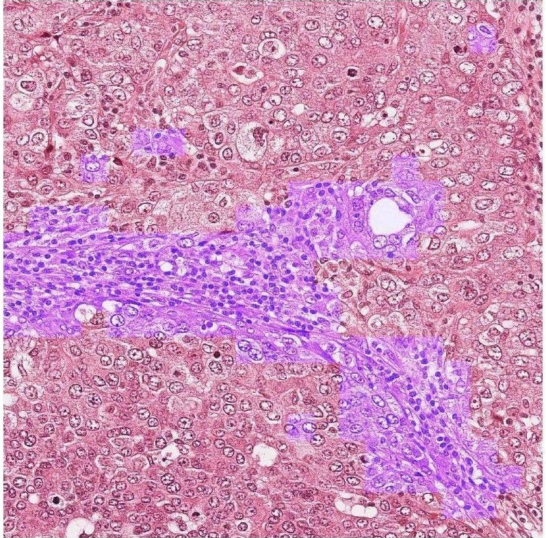
cancer. In designing a pipeline for the detection problem, identification of stroma in the tissue is
important when comes to rejection of false candidates.
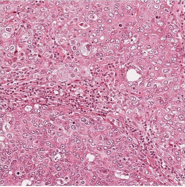
H&E stained breast biopsy tissue,
Results of stroma segmentation
|
|
| Medical Image Processing, CVIT, IIIT, Hyderabad
|
| Last Modified: Oct 31 2018
|
|
|