|
|
3D Retinal Image Analysis | |
Glaucoma, retinal vascular diseases, essentially neovascular age-related macular degeneration (AMD), retinal vein occlusion (RVO) and diabetic retinopathy(DR) are the leading causes of blindness in developed countries. Therefore, there have been efforts worldwide aiming at directly assessing the primary, structural consequences of disease activity using a new diagnostic method of retinal imaging, optical coherence tomography (OCT).
Following are some of the research projects of our group in the field.
- Segmentation of retinal cysts from SD-OCT scans
Retinal cysts are prominent indicators of eye diseases, thus their detection and segmentation is beneficial to clinical disease analysis and treatment. A most important ancillary test for the diagnosis of sight degrading diseases today is performed using spectral domain optical coherence tomography (SD-OCT). SD-OCT is a non-invasive modality for acquiring high resolution, 3D cross sectional volumetric images of the retina and the sub-retinal layers, in addition to retinal pathology.
We are working on a new novel automatic vendor independent retinal cyst segmentation methods. The dataset is collected from the OPTIMA Cyst segmentation challenge hosted at the MICCAI 2015 conference. Preliminary results was submitted to the challenge.
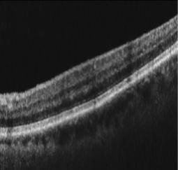
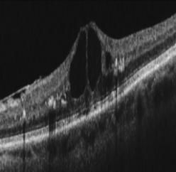 (L)Sample slice from an oct scanner. (R)Sample slice in presense of cyst
- Retinal Layer Segmentation from OCT Scans
Retinal Optical Coherence Tomography provide a cross-sectional view of intra-retinal tissue layers in a non-invasive manner. Accurate segmentation of these layers aids in computing layer thickness maps, tracking the progression of morphological changes in tissue layers over time and defining a region of interest for automated detection of pathologies for the clinical assesment of ocular diseases such as glaucoma and Age related Macular Degeneration (AMD). While spectral domain OCTs allow aquisition of large number of B-scans, their manual segmentation is tedius, time-consuming and subjective. Currently, commercial OCT systems are equipped to segment only two or three layers. The key challenges to segmentation are a) low signal-to-noise ratio due to speckle noise b) vessel shadows leading to intra-layer inhomogeneity c) lack of distinct boundaries and d) morphological changes in boundary smoothness.
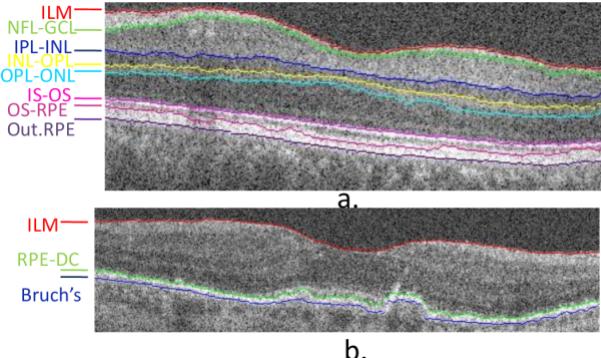 OCT layer Segmentation
- Glaucoma Classification from Angio-OCT images
OCT angiography (OCTA) is a new imaging modality providing angioflow profiles at various layers of the retina. It is based on a rapid OCT scanning of the eye at the same location
in the retina, over a time interval, to look for changes in the scan. All the structures in the retina are static except for the blood flow through vasculature or by movement of the eye
itself. Mapping these areas of blood flow is done using the Split-Spectrum Amplitude-Decorrelation Angiography algorithm to derive highly detailed maps of the vasculature in a noninvasive manner as opposed to fluorescein angiography. Specifically, in addition to structural information about the retinal layers, OCTA also provides angioflow (en face or top view) images which capture angiographic information at different layers namely, the superficial capillary plexus, deep capillary plexus, outer retina and choriocapillaris area which aid a detailed study of the capillary network.
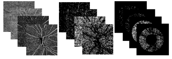 (L) DWI image, (R) Segmented Stroke
- Multimodal Registration in presence of Abnormalities
Image is more informative if two modalities are combined. On using OCT we can quantitatively know a variety of eye diseases like Localization of macular hemorrhage, qualitative and quantitative analysis of the optical nerve head and retinal nerve ber layer and analyzing progression of glaucoma. Exact localisation and morphology of abnormalities can be observed like drusen, macular hemorrhage. After registration we note there is more vessel information. Since some of the vessels appear in fundus image whereas some other vessels are better visualized in OCT images. This is in agreement with a paper that reports OCT projection images allow not only non-invasive visualisation of a 2D retinal perfusion map but also volumetric morphology of retinal microvasculature with high sensitivity.
Since some abnormalities are better visualised in OCT imaging like drusen, registration of two modalities helps to integrate all information in one picture. After registration of en-face with fundus. We get the transformation function. This can be applied to OCT en-face as different layer and original en-face can be replaced by en-face at different layer. By this we can visualize the vessels along with capillary vessels at different layers (for ex: like choroid vessels) on fundus image itself.
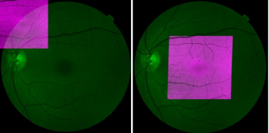 OCT en face in pink and fundus in green. Left image before registration. Right image after registration
|
|
| Medical Image Processing, CVIT, IIIT, Hyderabad
|
| Last Modified: Oct 31 2018
|
|
|