AnaVu: A practical 3D visualization system for integrative teaching of radiological and medical gross anatomy in large classrooms
We present AnaVu, a lightweight visualization system for teaching 3D anatomy at a classroom scale. We propose a stereoscopic system along with an easy-to-use interface as a scalable 3D visual aid as opposed to learning from traditional 2D images. This is an alternative to VR/XR devices that can only serve a handful of students and are heavy on computational resources. For large-scale classes (50-150 students), 3D visualization provides good feedback on spatial relations, with stereoscopic projection further providing depth cues to distinguish fine structures. The visualization is controllable by the lecturer with the ability to control interactive operators along with labels, animations and multimedia capabilities. Lessons can be premeditated and loaded quickly in class to integrate with the ongoing lecture.
A quantitative evaluation in two medical schools, involving 43 and 137 students, respectively, yielded results which show the proposed solution to be viable and effective for learning. AnaVu's integration of radiological anatomy enhances its applicability, providing students with a comprehensive understanding of anatomical structures in correlation with radiological images.
The main contributions of AnaVu are :
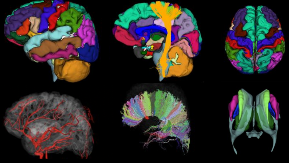
1. Structure extraction from in vivo images for precise rendition and registration of various 3D structures in volume and 2D slices.
2. A light-weight SceneGraph implementation supporting NIfTI, tractography, extracted 3D meshes, volumetric segmentation and volume files, animations and fluid simulations along with multimedia formats to show labels, videos and images.
3. A flexible GUI design - created with teacher inputs to keep various functions user-friendly for easy viewing and manipulation of on-screen data with support for 2D/3D switching and free focus on 3D data or volumetric slices for better contextual learning.