A diffeomorphic aging model for adult human brain from cross-sectional data
Normative aging trends of the brain can serve as an important reference in the assessment of neurological structural disorders. Such models are typically developed from longitudinal brain image data—follow-up data of the same subject over different time points. In practice, obtaining such longitudinal data is difficult.
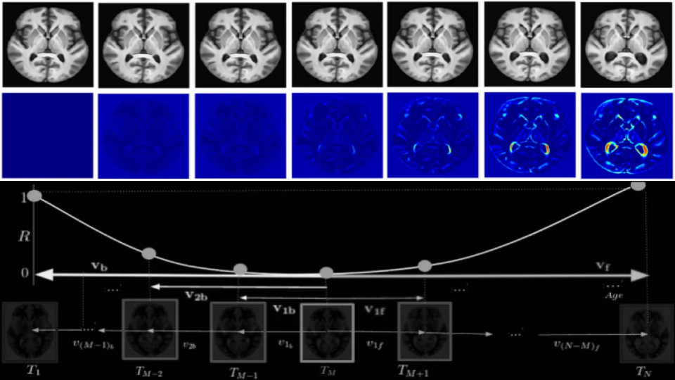
We propose a method to develop an aging model for a given population, in the absence of longitudinal data, by using images from different subjects at different time points, the so-called cross-sectional data. We define an aging model as a diffeomorphic deformation on a structural template derived from the data and propose a method that develops topology preserving aging model close to natural aging. The proposed model is successfully validated on two public cross-sectional datasets which provide templates constructed from different sets of subjects at different age points.
Understanding population-specific structural differences in brain aging
Understanding distinct neurological aging patterns across various populations is vital in the context of a globally aging populace. This study seeks to unravel the structural variations in the aging brain, taking into consideration different ethnic backgrounds, MRI data from Indian, Chinese, Japanese, and Caucasian populations were analyzed using a two-pronged approach.
Initially, a group analysis was performed involving tissue segmentation through FSL-FAST, examining gray matter (GM), white matter (WM), and cerebrospinal fluid (CSF). Subsequently, a continuous model-based analysis was employed, defining aging as a diffeomorphic transformation, which facilitated a detailed intra- and inter-population analysis, and examined both global anatomy and age-dependent distances of each population in comparison to the Indian population.
The analysis revealed distinct aging trajectories in the different populations. Notably, the Japanese demographic exhibited a delayed onset of GM reductions and ventricular expansion. Additionally, we observed a significant hemispherical asymmetry in the expansion rate in the left brain’s CSF-filled region across all populations.
Detailed insights into the spatial distribution of brain deformations over time were obtained, with particular focus on anatomical changes relative to a reference time point. Our findings are a step towards a nuanced understanding of the aging process, opening avenues for personalized age-related healthcare strategies rooted in a understanding of population-specific neurological aging trajectories.