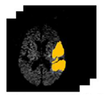
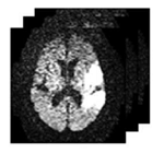
Detection and Segmentation of Stroke Lesions from Diffusion Weighted MRI Data of the Brain.
Shashank Mujumdar (homepage)
Stroke is a chronic disease which often leads to death. Different medical imaging modalities enable diagnosis for stroke after the onset of symptoms. Time is of the essence during stroke analysis since the window of therapy is very small (< 3 hrs after the onset of symptoms). Recent clinical studies have shown the usefulness and significance of diagnosing stroke on the Diffusion Weighted Magnetic Resonance Imaging (DWI) scans of the brain in the early stages. Visual inspection of the DWI scans is difficult since multiple scans are acquired for a patient with varied contrast and the scans depict complementary information about the diffusion process in the brain. To make matters worse, the DWI scans are acquired at a very low resolution with poor signal to noise ratio (SNR) since the time of acquisition is significantly less (< 1 min) and are confounded by artifacts that mimic stroke lesions. Thus, an automated framework which can accurately capture the stroke lesions in the DWI data would assist the clinicians in a better diagnosis. This is focus of the thesis.

Varying the acquisition parameter (b-value) generates different DWI scans with varied contrast. DWI with higher b-values provide improved sensitivity, conspicuity of stroke lesions and reduced artifacts at the cost of lower SNR. Along with the DWI scans, the Apparent Diffusion Coefficients (ADC) maps are also derived which give a measure of the true diffusion process in the brain irrespective of the acquisition artifacts that resemble stroke. In this thesis, we argue that integrating information from multiple sources, namely, low and high b-value data along with the ADC maps, can aid better characterization of stroke lesions in the data. Accordingly, we propose a novel approach for detecting and segmenting stroke regions from DWI data.
(more...)
| Year of completion: | July 2013 |
| Advisor : | Jayanthi Sivaswamy |


