Brain Image Analysis
Brain is one of the most complex and sophisticated organs in human body. It is the center of nervous system and responsible for motor actions, memory and intelligence in humans. Study of structure, function and disease in human brain is done by analyzing images obtained in different modalities like Magnetic Resonance Imaging (MRI) and Computed Tomography (CT).
Structural Analysis and Disease Detection in CT and MRI Brain Images
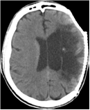
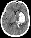
Stroke detection - Stroke is a disease which affects vessels that supply blood to the brain. A stroke occurs when a blood vessel either bursts or there is a blockage of the blood vessel. Due to lack of oxygen, nerve cells in the affected brain area are not able to perform basic functions and cause sudden death.
Strokes are mainly classified in two categories:
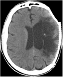
- Ischemic stroke or infarct (due to lack of blood supply)
- Hemorrhagic stroke (due to rupture of blood vessel)
 |
 |
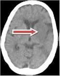
Computed tomographic (CT) images are widely used in the diagnosis of stroke. We are working on an automated method to detect and classify an abnormality into acute infarct, chronic infarct and hemorrhage at the slice level of non-contrast CT images. CT imaging is preferred over MRI due to wider availability, lower cost and lower scan time.
 |
 |
We have developed a unified method to detect both types of strokes from a given CT volume data. The proposed method is based on the observation that stroke presence disturbs the natural contra-lateral symmetry of a slice. Accordingly, we characterize stroke as a distortion between the two halves of the brain in terms of tissue density and texture distribution.
This work was conceived recently and is still under progress in association with CARE hospital.
Detection of Neurodegenerative diseases - Neurodegenerative diseases result in cognitive impairment that affects memory, attention, language and problem solving. It is caused due to head injury, infections or aging. Early detection of these diseases can be helpful in treatment and hence in reversing the impairment. MR images are recommended for detection of this class of diseases due to their high contrast property.
Structural imaging can detect and follow the time course of subtle brain atrophy as a surrogate marker for pathological processes. Our research is directed towards developing MR image analysis algorithms to detect such atrophies. Such detection tools will aid in studying the pathological processes and detect pre-demented conditions.
People Involved
- Sushma
- Rohit
- Shashank
- Mayank
- Sandeep
- Saurabh
